An Imitation "Melo Pearl"
January 21, 2011
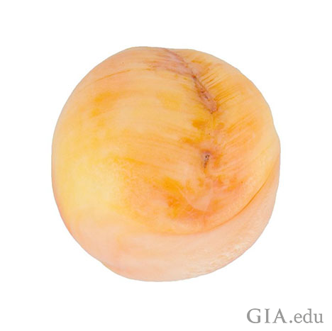
Every now and then a client will submit a “pearl” for identification that they have been told, or believe, is from a particular mollusk. Such was the case with the 52.49 carat near-round “pearl” measuring 19.62 x 19.21 x 19.16 mm, in figure 1 that was recently submitted as a Melo “pearl”. Whilst not an obvious pearl from its external appearance it certainly could pass as one, so more detailed examination, and therefore a report, was certainly warranted, and it was understandable why the client wanted to be certain of the result.
Preliminary visual examination with a loupe and subsequently a microscope revealed distinct relatively fine banding (figure 2) that formed a herring-bone pattern along a clear surface groove in one area. This in itself started to cause concern since it is very unusual to observe any structural banding in a pearl that can be considered “straight” unless it is somehow associated with the base of a blister pearl, when it might just be possible to get some straighter banding if there is any associated shell somehow still attached to the pearl. However, since the banding in this object appeared to run throughout the whole piece its “pearly” nature was immediately in doubt. To add even more doubt to the identity we also

noted that the color was a little more patchy than we would expect for a naturally colored pearl of this type, so closer examination was applied and it wasn’t long before some small but distinct orange color concentrations were observed within surface reaching cracks (figure 3) running off the main surface groove already described. This provided further evidence that all was not quite as it appeared. Yet we still had to gather more information to support our preliminary opinion and this meant that we had to turn to other equipment, including some of the more advanced analytical procedures, at our disposal.

After obtaining a specific gravity result of 2.84, we proceeded with our analysis by checking whether we could see any indications of a pearl-like structure using our in-house Faxitron real-time microradiographic system. This soon revealed a banded structure in the first orientation (figure 4) and a repeat of the herring-bone structure seen via the preliminary optical examination when it was first inspected, in another orientation. From our experience most natural Melo pearls reveal one of two microradiographic structures; either very little to no useful structure; or sometimes a small void-like feature near their centers. The banded nature of this particular sample was therefore not something we would associate with a natural concentrically formed Melo pearl, or pearl from any mollusk for that matter. The nature of the material forming this particular “pearl” was therefore concluded to be shell, so it appeared as though we were handling an example of an imitation Melo “pearl” fashioned from shell.

What about the color? We still had to prove whether it was naturally colored or not. Examination under ultra-violet lighting did not provide any useful reactions for either shortwave or long-wave radiations. We therefore focused on three pieces of equipment to help determine the color origin. Analysis with an energy-dispersive X-ray fluorescence spectrometer (EDXRF) revealed nothing out of the ordinary using a general method and confirmed that the shell used was fashioned from a saltwater mollusk owing to its low manganese and higher strontium content when a specific carbonate method was applied. A spectrum obtained after using a Perkin-Elmer 950 ultra-violet visible near-infrared spectrophotometer (UV-Vis-NIR) showed absorption features expected for an orange colored pearl or shell, with no features that would make us comfortable in making a treated color call, so this left us with the option of using a Renishaw inVia Raman microscope fitted with a 514 nm Ar-ion laser to determine the cause of color more specifically. The results of this analysis showed that the shell was composed of aragonite owing to the presence of one main vibrational peak at 1086 cm-1 and accompanying doublet at around 704 & 702 cm-1, but it also proved it was not naturally colored, since polyenic (Salares, 1977) peaks expected for naturally colored pearls or shell belonging to species such as Melo or Cassis amongst others, were lacking (Figures 5 and 6). Even in relatively lightly colored pearls with natural polyenic pigments some small peaks in the regions of 1120 cm-1 & 1520 cm-1 should usually be visible, so when the quite saturated colored areas of this “pearl” were examined it should have revealed these peaks quite strongly too. The fact that the peaks did not appear at all just confirmed that the coloration must be artificial.


Whilst we could not be more specific on the exact nature of the pigmentation used to color the shell, it bore a similarity to the dye detected in another imitation pearl submitted to the AGTA laboratory in 20081.
Another feature about the imitation that made its non-pearl status more likely was that it showed clear evidence of having been shaped and polished. Whilst heavy working (i.e. filing or grinding to alter the shape) and polishing to improve the luster are not proof in themselves of a shell origin, both of these actions are usually required to produce an imitation pearl from a piece of shell and so it makes it that much more suspicious when both are also present in addition to all the other factors that have been taken into account.
Dyed imitation Melo “pearls” are not a particular rarity in the market, but they do provide gemologists in laboratories something different to look at since they aren’t submitted on a particularly regular basis because most clients have a good idea whether they have an original pearl or not, and justifying the costs of testing something they suspect is an imitation is rather difficult. Therefore the only imitation pearls laboratories tend to see are those that clients truly believe are pearls, or ones that are submitted for the sake of interest/research (Krzemnicki, 2006; Wentzell, 2006, ). As with all of the imitation Melo “pearls” mentioned in the two articles referenced above, the most likely origin of this particular dyed shell is also the Tridacna species (various species of Clam including Tridacna gigas) based on the thickness of the shell and obvious flame pattern.
1 The article is no longer available via the AGTA website, but searches via the www can find a few references.
Krzemnicki, M.S., (2006), A Worked Shell Bead as an Imitation of a Melo Pearl
Salares, V.R., Young, N. M., Carey, P. R., Bernstein, H. J., (1977), Excited state (excitation) interactions in polyene aggregates. Resonance Raman and absorption spectroscopic evidence., J. Raman Spectrosc., 6, 282-288.
Wentzell, C.Y., (2006), Imitation Melo "Pearls", Gems & Gemology, 42, 2, 166-167.


